
Kili Technology
The data-centric AI platform for high-quality training data and model evaluation.
Updated 13d ago
Has API
PricingFree
Free
Image Segmentation
Named Entity Recognition (NER)
RLHF for LLMs

World’s leading Interactive Microscopy Image Analysis software for 3D and 4D imaging.

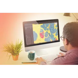
Imaris is a microscopy image analysis software designed for 3D and 4D datasets. It provides tools for visualization, segmentation, and quantification of microscopic images. The software architecture is built to handle large datasets efficiently, leveraging GPU acceleration for rendering and processing. Key value propositions include interactive analysis, AI-powered segmentation, and customizable workflows. Use cases span cancer research, cell biology, developmental biology, and neuroscience, allowing researchers to analyze cellular structures, track cell movements, and quantify biological processes. Imaris also supports integration with various microscopy systems and data formats, enabling seamless data import and analysis.
Imaris is a microscopy image analysis software designed for 3D and 4D datasets.
Explore all tools that specialize in segment images. This domain focus ensures Imaris delivers optimized results for this specific requirement.
Explore all tools that specialize in image segmentation. This domain focus ensures Imaris delivers optimized results for this specific requirement.
Utilizes deep learning algorithms to automatically segment complex structures in 3D/4D images, reducing manual effort and improving accuracy.
Provides interactive tools for visualizing large microscopy datasets, including volume rendering, surface rendering, and clipping planes.
Automated tracking algorithms to track cells and other objects over time in 4D datasets, enabling the study of cell migration and dynamics.
Quantifies the degree of overlap between different fluorescent labels, providing insights into protein interactions and cellular processes.
Allows users to automate image analysis workflows by processing multiple datasets simultaneously, improving efficiency and throughput.
Module designed for stitching multiple microscopy tiles into a single, large image. Useful for large tissue sections or whole-organ imaging.
Install Imaris software on a compatible workstation.
Import microscopy image data in supported formats (TIFF, LSM, VSI).
Use interactive tools to visualize 3D/4D datasets.
Apply segmentation algorithms to identify objects of interest.
Track objects over time using automated tracking algorithms.
Quantify object properties and generate statistical reports.
Customize workflows using Python scripting for advanced analysis.
All Set
Ready to go
Verified feedback from other users.
"Imaris is highly regarded for its advanced 3D/4D image analysis capabilities and user-friendly interface, though some users find it expensive."
Post questions, share tips, and help other users.
The data-centric AI platform for high-quality training data and model evaluation.
Discover and deploy pre-trained AI models for fashion-related tasks.
Vision Transformer and MLP-Mixer architectures for image recognition and processing.

High-performance computer vision framework for fashion analytics and virtual try-ons optimized for Huawei Ascend architecture.
Scikit-image is a Python package providing a collection of algorithms for image processing.
Trainable AI for insightful and robust image analysis in pathology.